A sensitive body part like your eyes need precise and sensitive care to ensure continued health. Dr. Edwin Schottenstein is committed to providing his patients with the expert eye care that they deserve with a compassionate approach. Dr. Schottenstein stays up to date with all of the most advanced ophthalmology procedures and technology to offer comprehensive ophthalmic treatment plans for even the most serious cases.
He proudly serves the areas of the Upper West Side, Washington Heights, Chelsea, and the greater Manhattan area out of his local office on West 71st Street between Broadway and Columbus Avenue.
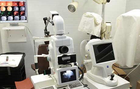
Below is a list of the ophthalmology technologies and methods used by Dr. Schottenstein to treat a wide array of eye conditions.
Digital fundus photography is a high-tech digital camera system used to take pictures of your retina. It has the capability of providing Dr. Schottenstein with more detail of the retina than other cameras can provide. These detailed pictures of the retina will help Dr. Schottenstein detect the presence of disease and discover eye problems not visible before digital fundus photography was available.
Digital fluorescein angiography is performed by injecting a special fluorescein dye into the arm. As it travels through the blood into the blood vessels in the back of the eye, a camera takes pictures of the eye. These pictures will detect any circulation problems, swelling, leaking or abnormal blood vessels.Unlike other angiogram procedures, an eye angiogram is not an X-ray procedure, so you are not exposed to any radiation.
Potential Acuity Meter (PAM) testing is used to assess retinal function. A Potential Acuity Meter is able to bypass cataracts and project an eye chart directly to the retina of the eye, allowing your doctor to test your vision without the interference of any cataract or problems with the lens of the eye. This is a particularly helpful test to perform on a patient who is considering cataract surgery.
he corneal specular microscope projects light onto the cornea and monitors the reflected light from an optical interface of the corneal tissue. Specular microscopes have been used most often to evaluate the corneal endothelium. However, with this reflective-light technology, the corneal epithelium, the stroma and the crystalline lens can all be examined and evaluated.
Optical Coherence Tomography is a noninvasive technique that is used by ophthalmologists to create ultra-high resolution, cross-sectional, detailed images of a patient’s retina. Optical Coherence Tomography is technology tool that has proven incredibly useful in evaluating the following conditions:
Perhaps the most compelling reason to use the new retinal nerve fiber layer / macular thickness topography imaging technology is that changes in the nerve fiber layer and optic disc are detectable by this technology years before they would otherwise be detectable in standard eye exam thus allowing for the earlier detection of a problem. These new advancements in technology have produced imaging technology that can more reliably measure change in disc size and shape and thinning of the nerve fiber layer, allowing for early detection. Early detection allows Dr. Schottenstein to intervene at the earliest possible stage of the disease’s progression and effectively monitor patients who are at a higher risk of developing such problems.
Anterior chamber angle (ACA) and anterior chamber depth (ACD) assessments are necessary in order to diagnose narrow angle and closed angle glaucoma as well as pigmentary glaucoma and neovascular glaucoma. Available methods of testing include:
Most of the tests mentioned previously can be carried out with the use of a slit lamp. A slit-lamp examination uses a microscope with a light attached, which allows the Dr. Schottenstein to examine the structures at the front of the eye (cornea, iris and lens) under high magnification.
A brightness acuity test uses low, medium and high light setting on the brightness acuity instrument to diagnose disability in three light conditions: direct overhead sunlight, a partly cloudy day and bright overhead lighting. If a patient’s vision worsens while being exposed to light from the brightness acuity testing instrument, the patient is considered to have a glare disability, typically caused by cataracts.
Corneal topography produces a detailed map of the shape of the cornea. Computerized imaging technology produces a 3D map that provides detailed images that help in diagnosing, monitoring and treating different conditions of the cornea. The shape of a person’s cornea has a lot to do with determining the visual capabilities of an otherwise healthy eye. A perfect eye has an evenly rounded cornea. If the cornea is too flat, too curvy, too steep or otherwise misshapen, there will be varying degrees of vision impairment.
Ultrasound technology uses high frequency sound waves to produce a two-dimensional view of eye tissue. The sound waves are reflected by eye tissues and orbital structures and converted into electrical pulses that are recorded on a printout. The most successful form of ultrasound uses the immersion method, during which the eye is in contact with a water bath for clearer images. The water bath technique provides better resolution than other ultrasound techniques.
The WHITESTAR Signature™ System is a revolutionary surgical breakthrough that allows the safe and easy removal of the lens, a technique that has previously not been available to cataract surgeons. This state-of-the-art system is extremely versatile and can safely remove both hard and soft lenses.
Accommodating IOL are lenses designed to adjust like the natural eye, so that a patient can see at multiple distances. Typically used in cataract surgery, accommodating lenses attempt to reduce the need for glasses after surgery.
Conventional IOL are only monofocal, which means that patients only have vision at one set distance (far, intermediate or near). Corrective lenses are still needed to compensate for the other distances that are not set through the conventional IOL. Now, there are new multifocal IOL available, which provide patients the ability to see well at more than one distance without the use of glasses or contacts. Some examples of multifocal IOL include:
The Humphrey Visual Field Analyzer will allow Dr. Schottenstein to see the full horizontal and vertical range of what you are able to see peripherally. They determine the potential of blind spots (scotomas) occurring, which could indicate eye diseases. During the test, you will be asked to cover one eye while focusing on a specific target object and asked to respond to certain light stimuli in your peripheral vision. If an eye disease is suspected, further comprehensive, more formal types of tests may be required.
The argon laser is used in the treatment of multiple eye disorders such as:
An argon laser uses argon gas to produce particular blue and green wavelengths that are absorbed into the eye without damage. Argon laser technology has the ability to seal blood vessels in the eye, reattach detached retinas, close tiny holes in the eye and destroy abnormal blood vessels.
The lens of the eye is positioned in an elastic-like capsular bag, which holds it in place and serves as a protective barrier. During cataract surgery, the front portion of the capsule in opened so the lens can be removed and replaced with an intraocular lens.
In some instances, a person may experience blurred, hazy vision, a common condition known as posterior capsular haze (sometimes referred to as “secondary cataract”). An estimated 40% of patients who have cataract surgery will experience posterior capsular haze. It can occur months or even years after the surgery. The YAG Laser is sometimes used by Dr. Schottenstein to treat this condition. This treatment is called YAG Laser Capsulotomy.
YAG Laser Capsulotomy is an outpatient procedure, which only takes a few minutes and is entirely painless. After the eyes have been dilated, Dr. Schottenstein will use the YAG Laser to create an opening in the center of the cloudy capsule. The majority of patients will notice an instant improvement in vision, while others will experience a gradual improvement over a period of days. Following the procedure, your vital signs and intraocular pressure will be reviewed. You can often return to normal activities immediately.
A refraction assessment helps Dr. Schottenstein determine the most accurate corrective lens prescription that will give you the best possible vision. The Canon Auto-Refractor is designed for easy use and the most accurate results. You will be asked to look through a Phoroptor, a mask-like device that contains different lenses, which will help determine the best combination that will give you the sharpest vision. The motorized optical head automatically aligns, acquires the readings and then transitions to the opposite eye to perform the same testing function.
Dr. Schottenstein is one a board certified ophthalmologists, and one of New York City’s best. He has had almost 30 years of experience practicing ophthalmology and helping patients just like you with their eye conditions, so you know that you will get the compassionate and professional eye care that you deserve. Issues with vision are something that should always be taken seriously, so if you an ophthalmologist, contact us and schedule an appointment with Dr. Schottenstein today. Him and his qualified staff are standing by to answer any questions and create the best treatment plan for you.